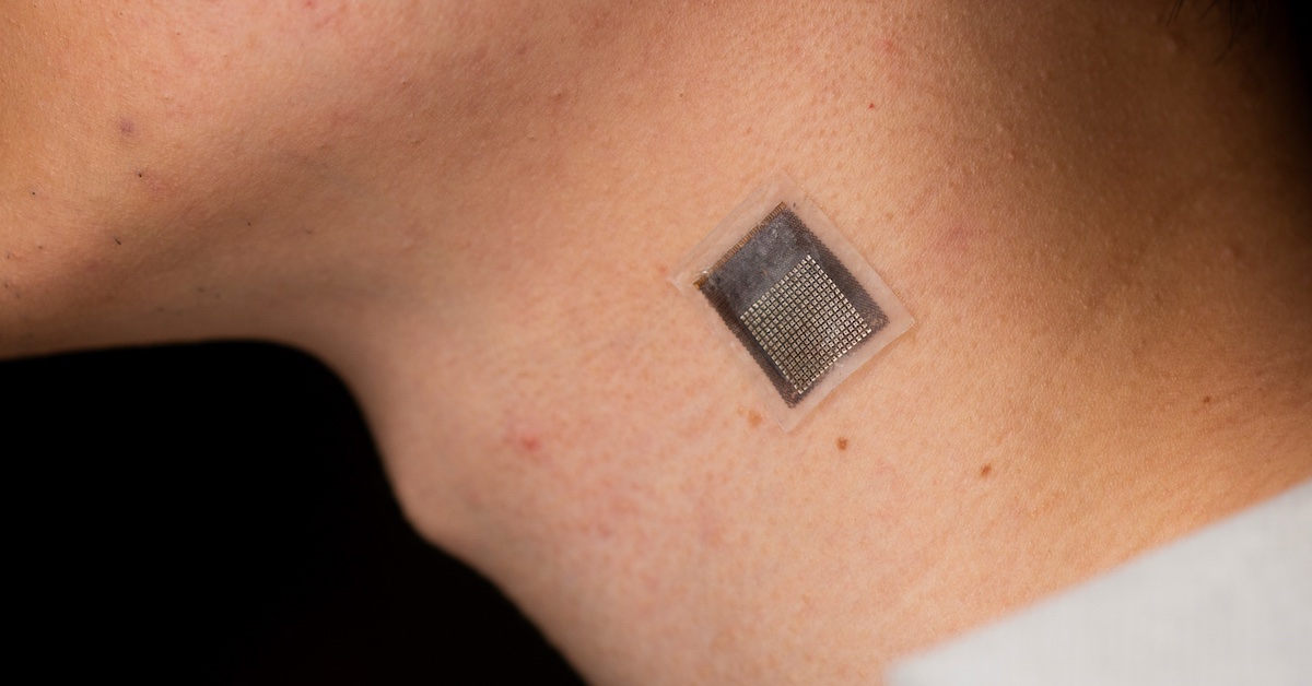
A team of engineers at the University of California San Diego has developed a new method that uses ultrasound to examine the biomechanical properties of tissues in a non-invasive manner. The method can help detect and manage pathophysiological conditions, track the evolution of lesions and evaluate the progress of rehabilitation. The stretchable ultrasonic array developed by the team facilitates serial, non-invasive, three-dimensional imaging of tissues up to four centimeters deep below the surface of human skin, at a spatial resolution of 0.5 millimeters. This is a significant improvement over current methods that have limited penetration depth.
“We invented a wearable device that can frequently evaluate the stiffness of human tissue,” said Hongjie Hu, a postdoctoral researcher in the Xu group and study coauthor. The team integrated an array of ultrasound elements into a soft elastomer matrix and used wavy serpentine stretchable electrodes to connect these elements, enabling the device to conform to human skin for serial assessment of tissue stiffness.
The new method has several key applications. In medical research, serial data on pathological tissues can provide crucial information on the progression of diseases such as cancer, which normally causes cells to stiffen. Monitoring muscles, tendons, and ligaments can help diagnose and treat sports injuries. Current treatments for liver and cardiovascular illnesses, along with some chemotherapy agents, may affect tissue stiffness. Continuous elastography could help assess the efficacy and delivery of these medications and aid in creating novel treatments.
Apart from monitoring cancerous tissues, the technology can also be applied in other scenarios such as monitoring fibrosis and cirrhosis of the liver, assessing musculoskeletal disorders such as tendonitis, tennis elbow and carpal tunnel syndrome, and diagnosing and monitoring for myocardial ischemia. By monitoring arterial wall elasticity, doctors can identify early signs of the condition and make timely interventions to prevent further damage.
The technology represents a significant advance in non-invasive, longer-term alternative methods for examining tissue stiffness. The researchers plan to further develop this technology to improve its capabilities for monitoring changes in tissue stiffness over time. The paper describing the research, “Stretchable ultrasonic arrays for the three-dimensional mapping of the modulus of deep tissue,” is published in the May 1, 2023 issue of Nature Biomedical Engineering.
Source: UC San Diego Today
